
CT Scans
Blue Cross Animal Hospital provides high-quality diagnostic imaging to assist with the state-of-the-art care and treatment of pets in our hospital. We offer computed tomography studies using an 8-slice Neurologica Ceretom CT scanner.
Computed tomographic images are obtained using X-rays, similar to normal radiographs, but the X-rays are generated and detected in a ring-like structure (the gantry) that surrounds the patient. The detected information is displayed as an image by assigning each pixel of data to a shade of gray.
Multi-detector (slice) CT scanners derive their name from the fact that there are multiple detectors available to receive the X-rays that have passed through the patient, giving us the ability to simultaneously obtain multiple cross-sectional images, compared with a single-detector unit. This speeds up the image acquisition process by a factor of the number of slices the machine can acquire. Most CT studies require general anesthesia but typically take less than 20 minutes to complete.
All of our studies are forwarded to a board-certified radiologist to interpret and we can have results as fast as the same day of the scan.
CT studies performed by Blue Cross include:
- Head and Neck
- Thorax
- Abdomen
- Spine
- Pelvis
Abdominal Ultrasound
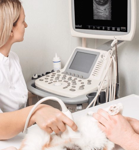
Blue Cross Animal Hospital offers state-of-the-art ultrasound imaging. We have over 25 years of experience in performing and interpreting ultrasound studies on companion animals. We have invested in state-of-the-art ultrasounds from Toshiba and Esaote. Our imaging studies are interpreted on site and full reports are provided within 72 hours. We communicate all of our findings with you as well as your referring veterinarian. Studies that we perform are:
- Abdominal ultrasound
- Porto-systemic shunt evaluation (must have bile acids prior to study)
- Full echocardiogram (right parasternal, left cranial and left caudal parasternal views, M Mode measurements, 2 D measurements, spectral and color flow Doppler evaluation)
- Cervical ultrasound of the thyroid and parathyroid
- A-FAST
- T-FAST
